INM’s World Leading Clinical Physics Team.
Clinical Physics at INM.
The Clinical Physics group is responsible for the provision of scientific support to the clinical Nuclear Medicine department and is also active in many research areas: the group lead on a number of research projects and support clinical and translational research within the Institute of Nuclear Medicine and beyond.
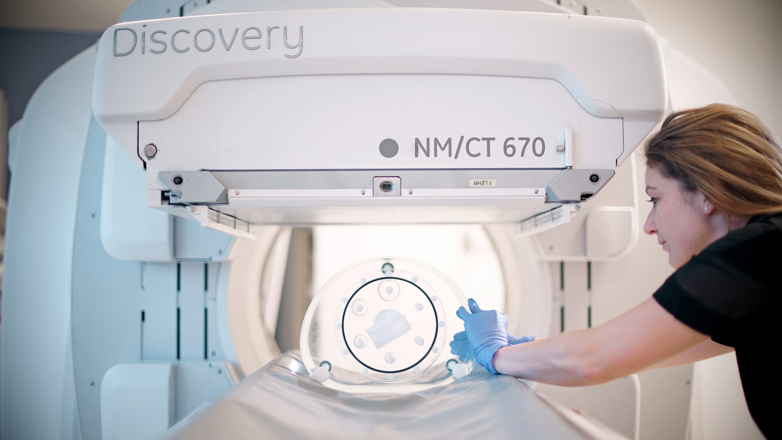
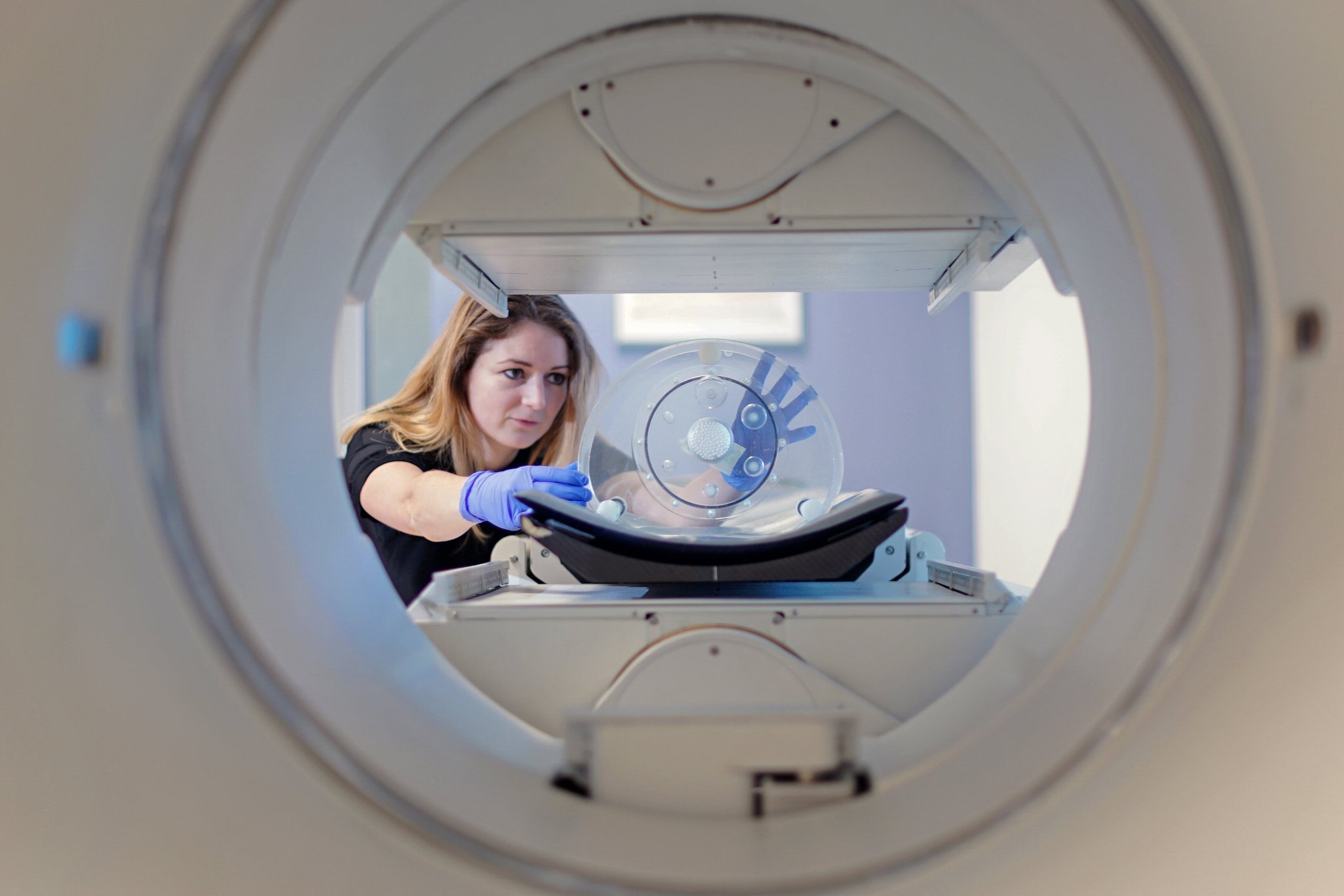
We support the clinical department in many ways; we help to ensure that the patient images acquired contain the best possible diagnostic information by optimising image acquisition and reconstruction, and performing regular quality assurance of the department’s state-of-the-art PET-CT, PET-MR and SPECT-CT imaging systems. We also play an important role in the development of the service provided to patients, through introducing and evaluating new techniques. We are central to the Molecular radiotherapy service; we are involved in a wide range of therapy administrations and we are working to implement and improve methods of dosimetry calculations, to provide patient-specific treatment information.
Quantitative SPECT.
SPECT images are conventionally interpreted qualitatively, which can be subjective and limits the reporting doctor’s ability to precisely compare changes in radiotracer uptake over time. The addition of accurate quantitative data could facilitate more objective assessments of disease extent, rendering diagnostic reports more comprehensive and enabling us to better monitor lesion progression and response to treatment.
We are investigating quantitative SPECT as a routine diagnostic tool; to determine how to overcome degrading effects such as the ‘Partial Volume Effect’ that can inhibit its accuracy, as well as assessing the clinical utility of introducing these measurements to the interpretation process.

PET Image Optimisation & Research.
We do a large amount of work in optimising image quality for each study type in Positron Emission Tomography (PET) imaging so as to get the best possible diagnostic information from each scan. With our state-of-the-art PET-CT systems, we are also able to investigate new technologies, such as parametric imaging, novel quality control techniques and respiratory gating methods, which will impact on the service we are able to provide to our patients.

Dosimetry for Molecular Radiotherapy.
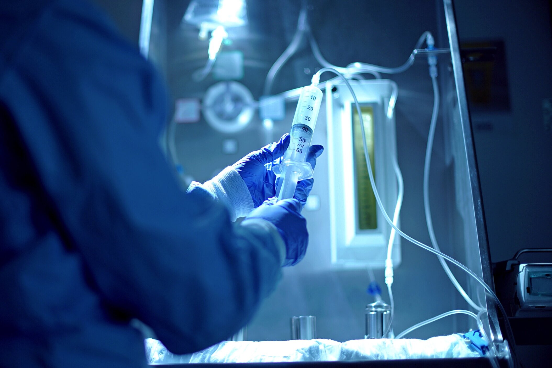
Many molecular radiotherapy cancer treatments are performed at UCLH and the clinical physics team are heavily involved in these; both in the clinical provision of the service and in optimising dosimetry techniques. The aim of dosimetry measurements is to calculate the radiation dose to both the target and to healthy organs and requires multiple SPECT-CT images to be acquired and processed (each organ needs to be outlined and the time-activity-curve in each organ analysed) in order to perform these calculations. The clinical physics group are involved in performing these calculations for clinical trials and are working towards improving the methodology for future patients.
Smith A L, Sanderson T et al “Internal Dosimetry considerations for paediatric radioimmunotherapy” BNMS Annual Meeting, 2017
Pre-surgical Epilepsy Imaging.
Imaging is used to guide epileptic surgical procedures that are carried out in order to reduce seizures and the impact they have on the patient’s quality of life. It is vital for surgeons to accurately identify the location within the brain that needs to be targeted during surgery and these ictal-interictal (during-seizure and between-seizure) studies provide this crucial information.
Our team are investigating how to optimise the information that can be extracted from these studies, for example using voxelwise statistical analysis, and are liaising with the telemetry team at NHNN to develop the service further and improve epilepsy diagnosis.
Smith A L et al “Experience at UCLH of Ictal and Interictal 99mTc-HMPAO SPECT/CT imaging in localising epileptic seizure site: a viable alternative to 18F-FDG PET/CT”, BNMS Annual Meeting, 2017

Movement Disorder Imaging .
Movement disorders such as Parkinson’s disease can be diagnosed with Nuclear Medicine imaging of [123I]FP-CIT, which binds to dopamine transporters. Quantification of these images provides invaluable information in their interpretation, but techniques for quantification are known to vary between centres.
The group have had a substantial input into studies investigating the factors that affect quantitative accuracy, highlighting the substantial impact of different imaging choices and serving as guidance in the production and interpretation of these measurements.
Dickson, J. C., et al (2017) “The impact of reconstruction and scanner characterisation on the diagnostic capability of a normal database for [123I]FP-CIT SPECT imaging” EJNMMI Research
Tossici-Bolt, L., Dickson, J. C., et al (2017) “[123I]FP-CIT ENC-DAT normal database: the impact of the reconstruction and quantification methods” EJNMMI Physics
Smith A L et al “Effects of different reconstruction methods on 123I-FP-CIT (DaTSCAN) SPECT quantification”, EANM Annual Meeting, 2017

Insight 46.
“Insight 46” is a neuroscience sub-study of the MRC National Survey of Health and Development. This is one of the oldest British birth cohort studies and has followed 5362 individuals since their birth in England, Scotland and Wales during one week in March 1946. Now aged 71 years, estimates suggest that ~1/3 of individuals in this age group may be in the preclinical stages of Alzheimer’s disease. This study involves a prospective two time-point data collection covering clinical, neuropsychological, β-amyloid positron emission tomography and magnetic resonance imaging, biomarker and genetic information.